
Review of imaging modalities of hip arthroplasty
In a paper published in France in the Journal D'imagerie Diagnostique et Interventionnelle, Pelissou et al. reviewed the imaging modalities of hip arthroplasty with a special focus on complications occurring in general, and specifically with respect to bearing couples. Knowing the possible complications related to THA helps the radiologist systematically to look for specific imaging patterns.
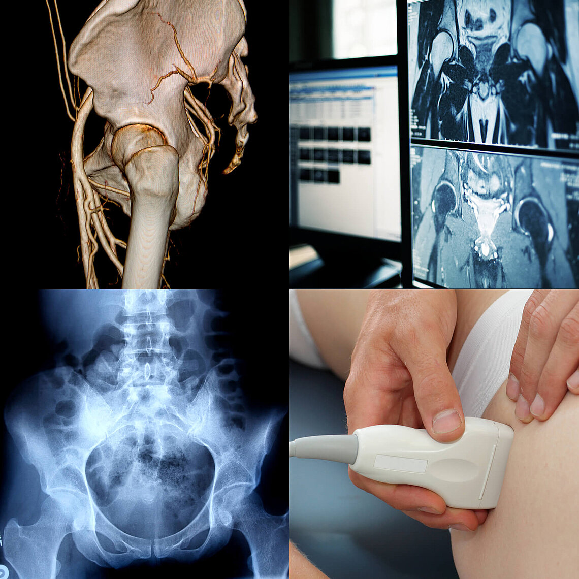
The conclusions:
- X-rays remains the routine examination for THA monitoring: Positioning, lysis, stress shielding, spotwelds, calcar
- CT scans are used for painful hips. It allows a fine analysis of bone/cement and bone/prosthesis interface. It helps better to analyse periprosthetic osteolysis, infra-radiological fractures (e.g., stem, ceramic) and to measure acetabular and femoral versions
- (MARS)-MRI, VAT and MAVRIC are particularly useful for soft tissue analysis (especially in the case of pseudotumors)
- Ultrasound depends on the echogenicity of the patient. It is preferably used for tendinopathies, psoas-capsular conflicts and puncture of periprosthetic collections
- Arthrography – not commonly used – is used to detect joint fluid collection in the event of loosening
Reference: Pelissou C, Miquel A, Phan C, Paycha F, Sautet A, Arrivé L. L’imagerie des prothèses de hanche: complications communes et spécifiques des différents couples de frottements - Imaging of hip arthroplasty: Common and specific complications of different bearing couples. Journal d'imagerie diagnostique et interventionnelle. 2020;3(1):47-66. doi:10.1016/j.jidi.2019.10.002.